Ambient AFM
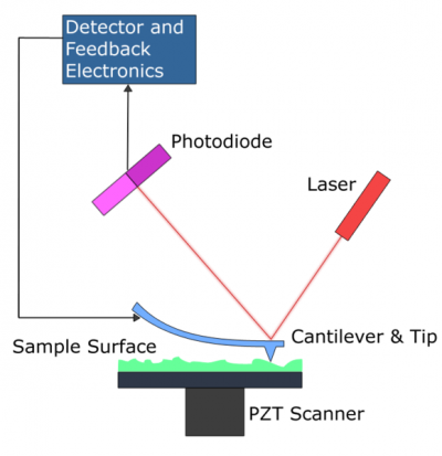
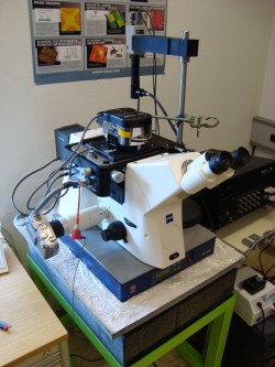
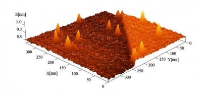
We are using an ASYLUM RESEARCH AFM to investigate surfaces at nanoscale in ambient conditions. The AFM is a very high resolution type of scanning probe microscope for imaging, measuring and manipulating matter. The AFM consists of a cantilever with a low spring constant, providing a sharp tip at its end which is used to scan the sample surface, an IR-laser aligned with the cantilever and a deflection sensor which is a four-quadrant-photodiode.
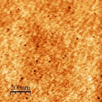
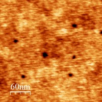
The tip of the cantilever is scanning the surface, reacting in response to the forces between the tip and the sample surface. A laser focused on the back of the cantilever and reflected onto a split photo-diode makes the smallest responses of the tip visible. By keeping track of changes in signal, the bending of the cantilever due to the interaction with the surface can be measured and the forces bending the cantilever can easily be derived, given a known spring constant of the cantilever. Obtaining data by this simple method, the image displays the surface in all three dimensions without the limit of optical diffraction in contrast to optical microscopes - thus resolutions several nm (in ambient conditions) or even atomic resolution (in UHV conditions) are possible.
Investigations can either be performed on any flat surface in air or in any fluid. Several operational modes are possible, the most common ones are CONTACT MODE and TAPPING MODE, enabling a wide approach for measuring various effects on surfaces - like roughness, local magnetic properties, defects induced by radiation or toughness. Since the cantilever is barely in contact with the surfaces, or even not even touching it at all (non-contact mode), the AFM provides a versatile, non destructive tool for investigation.
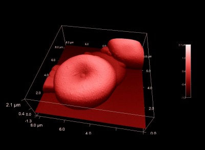
The main advantage of the ambient AFM is the simple setup for measurements - a sample is set up within minutes, cantilever changes are just as easy. Thus even living bacteria (like bacillus subtilis), human blood cells or chemical reactions on surfaces have been investigated.
For further information please contact:
* Gösselsberger Christoph ([email address: lastname @ this server · enable javascript to see it])
* Ritter Robert ([email address: lastname @ this server · enable javascript to see it])
* Vasko Christopher ([email address: lastname @ this server · enable javascript to see it])


