Scanning Auger Microscope - Focused Ion Beam
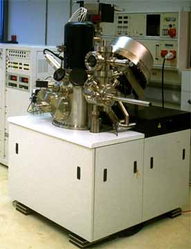
Our Scanning Auger System (VG microlab 310F) feature high spatial
and spectral resolution, due to it's field emission electron source
and its hemispherical electron analyzer with retarding lens. the
lateral resolution determined by the focus of the electron column
is 10...20 nm, while the depth resolution of 0,5...2 nm is
determined by the inelastic mean free path of the signal electrons.
The energy resolution is better than 1 eV. Primary energies of 50
eV to 25 keV can be used. The system is equipped with a sputter ion
gun (EX 05, Ar ions of 0,5 to 5 keV) for depth profiling, a heating
stage to 600 °C, a fracture stage and a focussed ion beam
column. A very similar new model is called
Microlab 350
The instrument is generally used for elemental analysis of various
objects on the micro and nano scale. It can detect all elements
with the exception of hydrogen and helium down to concentrations of
1 atomic percent. All electrically conductive samples and many
insulators can be analyzed with lateral resolutions down to 50 or
100 nm. Achieving the top resolution requires well prepared
conductive samples.
Besides general analytic applications, the instrument is used for
electron transport studies, e.g. to derive information used for
SESSA
The ion gun is used to clean surfaces and to record depth profiles
ranging from a few to several 100 nm. Larger depths can be analyzed
using either the FIB-column (to 10 μm) or special preparation techniques.
Caption for picture.
The focussed ion beam column (FIB) permits
micro-machining of samples in situ. This can be used for a variety
of purposes, e.g. the analyses of thick layers in the micrometer
range or of 3-dimensional micro- and nano-structures.
The fracture stage exposes internal surfaces of samples under high
vacuum. A typical application is the detection of impurities in
grain boundaries.
The heating stage is used to observe the effect of heating of
samples up to 600 °C on-line.
Sample requirements: Sample sizes can range from below 1 mm up to
38 mm diameter and 8 mm thickness. Up to a size of 10 mm by 10 mm
and a thickness of 3 mm every spot of the sample surface is
accessible. For the fracture stage elongated samples up to 30 mm
length and 5 mm diameter are used. Consult us for smaller samples
for the fracture stage and for samples for the heating stage. We
are able to perform a variety of preparation
tasks. Important: Samples must have a very low vapor pressure
and must not decompose under intense electron irradiation (Contact
us when in doubt).
Please direct inquiries for analytical work to Herbert Störi or Christian Tomastik.
